-
-
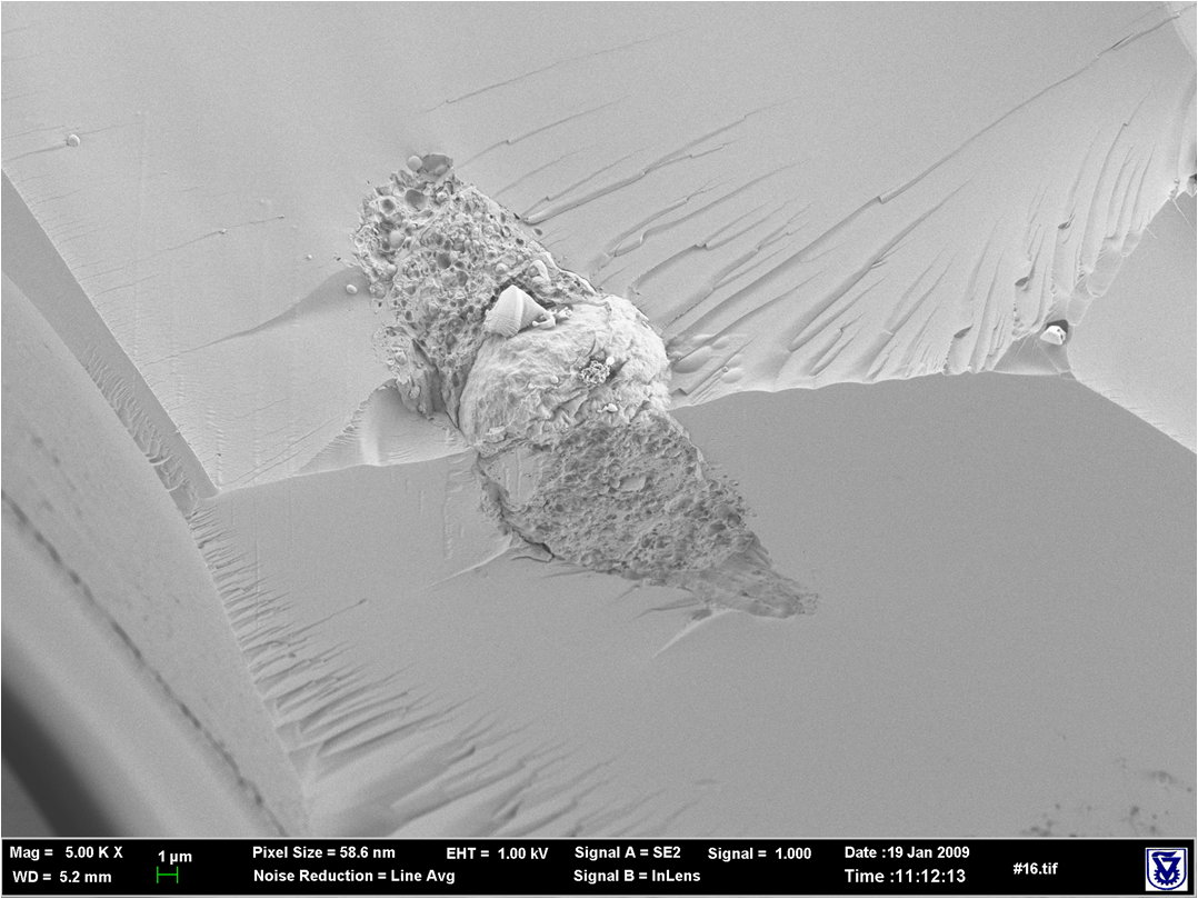
Cory-SEM image of a mesenchymal cell encapsulated in a PEG-fibrinogen hydrogel after several days in 3-D culture
-
-
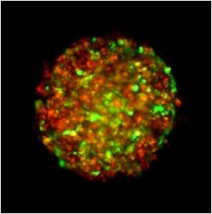
“Gel-in-gel” co-cultures of metastatic (red) and mesenchymal cells (green)
-
-
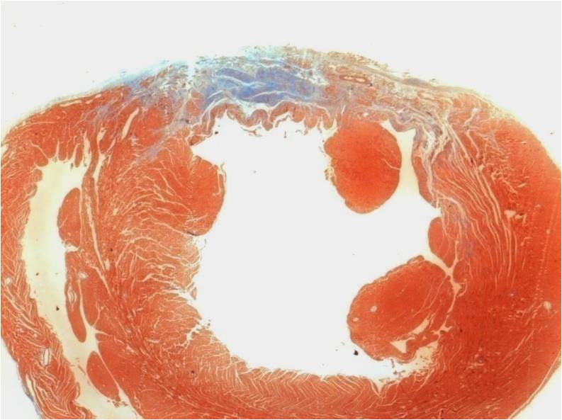
Cardiac myocytes injected into a myocardial infarction in a rat heart using a PEG-fibrinogen cell carrier; wall thickness after 30 days
-
-
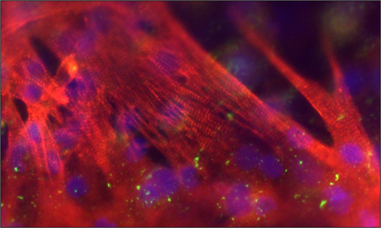
Cardiac myocytes cultured within PEG-fibrinogen hydrogels after several days; staining for α-Sarcomeric actin, Cx43 and DAPI
-
-
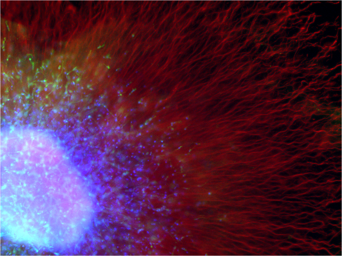
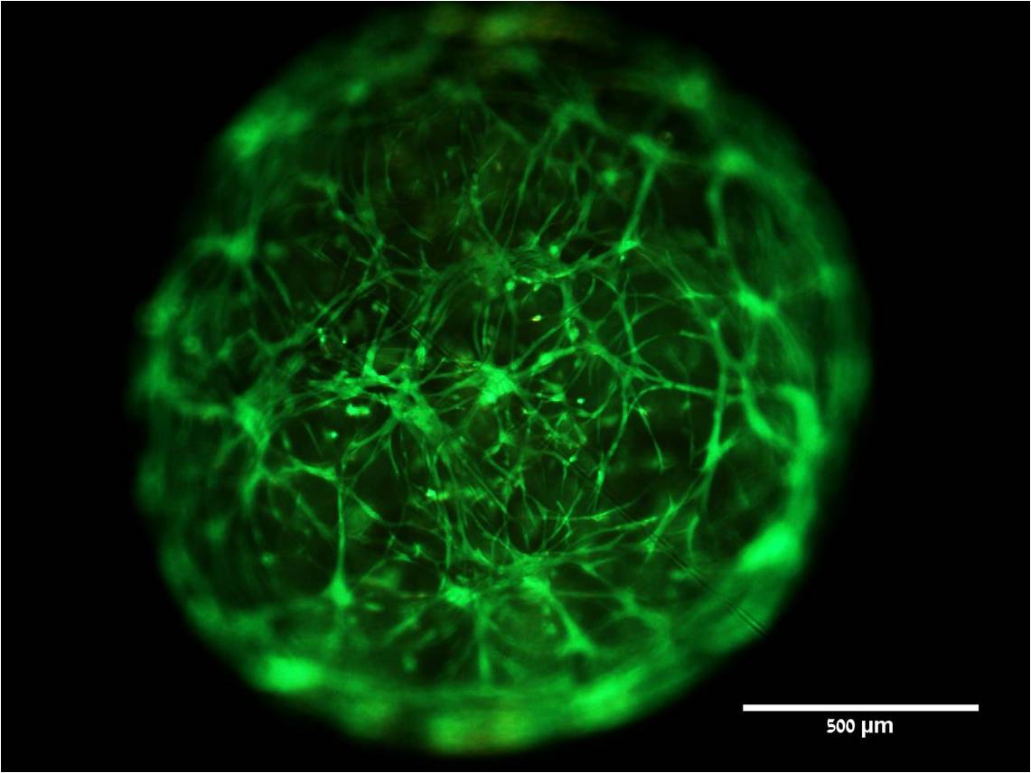
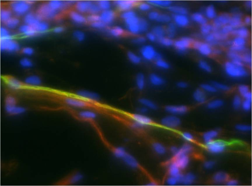
Dorsal root ganglion outgrowth into PEG-fibrinogen hydrogels after 1 week in culture; shown are neuronal cells (red) and glial cells (green)
-
-
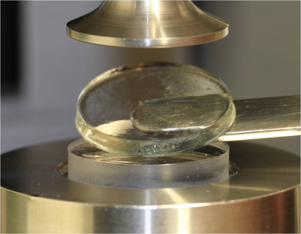
Parallel plate rheometry measurements performed on PEG-fibrinogen hydrogels with UV-curing attachment
-
-
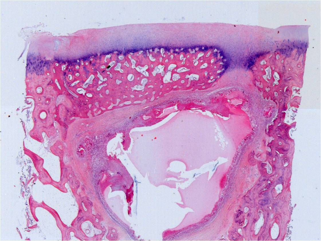
PEG-fibrinogen hydrogels used in the regeneration of osteochondral defects in sheep; new bone and cartilage visible
-
-
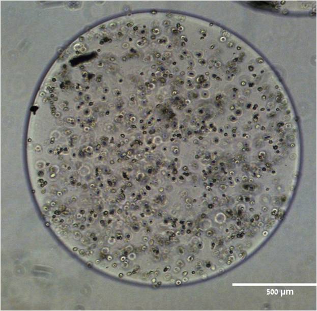
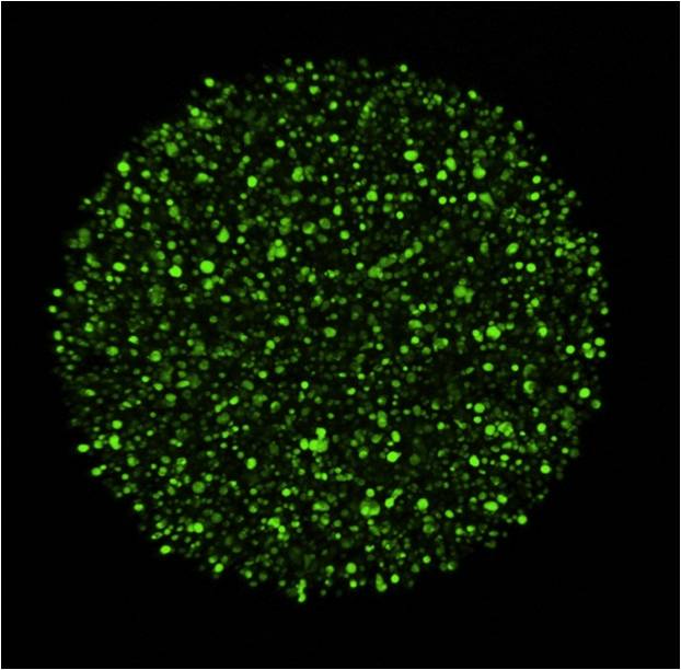
PEG-fibrinogen microgel culture of GFP-labeled mesenchymal cells immediately after encapsulation
-
-
PEG-fibrinogen microgel culture of GFP-labeled mesenchymal cells immediately after encapsulation
-
-
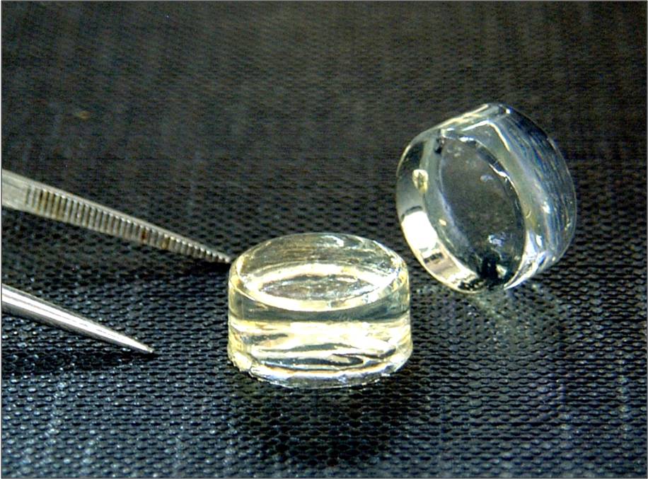
Transparent hydrogels made from PEG-fibrinogen containing cells are cast into the shape of disks for studying cells in 3-D culture
-

-
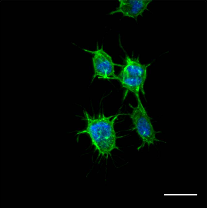
Mesenchymal cell spreading within PEG-fibrinogen hydrogels after 7 day in culture.
-
-
Mesenchymal cell spreading within PEG-fibrinogen hydrogels after 1 day in culture.
-
-
PEG-fibrinogen microgel culture of GFP-labeled mesenchymal cells 6 days after encapsulation
-
-
Dorsal root ganglion outgrowth into PEG-fibrinogen hydrogels after 1 week in culture; shown are neuronal cells (red) and glial cells (green)
-
-
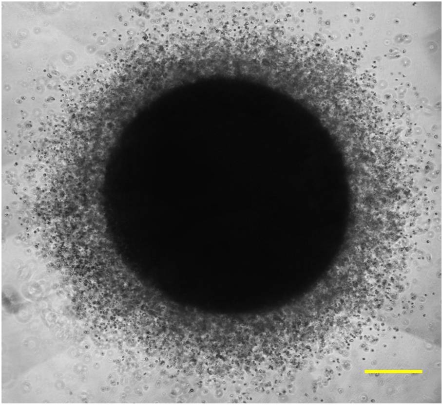
“Gel-in-gel” co-cultures of metastatic and mesenchymal cells; metastatic cell outgrowth visualized by phase contrast microscopy